Steric stabilizers inhibit or enhance enzymatic digestion depending on the internal nanostructure of lipid nanoparticles
Jonathan Caukwell, Karl A. Hassan, Brett A. Neilan, Andrew J.Clulow, Benjamin Boyd,Wye-Khay Fong and Livia Salvati Manni
This article is reposted with kind permission from The Royal Society Publishing. Original publication: Interface Focus, Vol. 16, Issue 2 (August 2026)
Link to article | https://doi.org/10.1098/rsfs.2026.0001
Abstract
Lipidic nanoparticles (LNPs), including liposomes and cubosomes, are widely employed drug delivery systems owing to their biocompatibility and capacity for stimuli-responsive ‘smart’ drug release. Steric stabilizers, including DSPE-PEG-2000 and Pluronic F127, are commonly used to stabilize LNPs by creating an effective steric barrier between the particle and aqueous physiological environment, but the implication for interactions with macromolecular components of complex physiological fluids is unclear. Thus, the impact of the polymeric steric barrier on enzymatic digestion by bacterial lipases is investigated in this study. Specifically, the impact of stabilizer chemistry, mode of attachment and nanoparticle geometry on the digestion of phospholipid liposomes and monoolein cubosomes by cell-free supernatants of Staphylococcus aureus and Pseudomonas aeruginosa containing the expressed extracellular enzymes from these organisms is investigated. Small-angle X-ray scattering (SAXS) revealed that bilayer-anchored DSPE-PEG-2000 hinders the digestion of phospholipid liposomes yet accelerates the digestion of monoolein cubosomes. These results suggest that nanoparticle architecture determines how effectively steric stabilizers protect against enzymatic digestion.
This article is part of the theme issue ‘Geometry, materials and the imagination’.
1. Introduction
Lipidic nanoparticles (LNPs) are a leading drug delivery system platform. Capable of carrying hydrophilic, hydrophobic, amphiphilic, small-molecule, peptide- and protein-based therapeutic agents [1,2], they provide a versatile means for expanding the range of therapeutic agents that can be used in medicine [3]. Phospholipid liposomes represent the most established and widely used class of LNPs [4–6], while monoolein cubosomes represent the most extensively studied non-lamellar LNPs [1,7]. The key feature that elevates these systems is their potential for ‘smart’ drug release—undergoing context-dependent lipid reorganization to different self-assembled structures which differ in release kinetics by orders of magnitude [8–11]. This stimuli-responsive release can be triggered by biological stimuli [12–14], while responsiveness can be tuned by particle composition [7,15]. Understanding the relationship between particle composition, biological stimuli and the ensuing nanostructural response is fundamental to unlocking ‘smart’ drug delivery with these systems.
Most LNPs require a steric stabilizer to maintain their stability and integrity in physiological fluids. Unstabilized LNPs are susceptible to aggregation, enzymatic digestion and recognition by the mononuclear phagocyte system (MPS), which clears them from the bloodstream [16]. Bilayer-anchored lipid-PEG conjugates such as DSPE-PEG-2000 and surface-adhering triblock copolymers such as Pluronic F127 are the most widely used LNP stabilizers [3,17]. DSPE-PEG-2000 features a hydrophilic polyethylene glycol (PEG) chain covalently bound to phospholipids in constituent lipid bilayers. It is typically employed for preventing protein adsorption to lamellar LNPs and for creating an attachment point for surface-functionalization with application-specific biomolecules [18]. Alternatively, Pluronic F127 features two hydrophilic PEG blocks connected via a hydrophobic polypropylene glycol (PPG) block that adheres to the bilayer surface and is typically employed for stabilizing non-lamellar LNPs [17]. Surface-adhering stabilizer molecules associated with the surface are in equilibrium with those in the surrounding solution, meaning their detachment is a potential mechanism for enabling biomolecular stimuli, such as enzymes, access to the LNP surface [19].
Although the stabilizer layer may partially and temporarily block the adsorption of proteins onto the surface of the particles, LNPs eventually interact with biomolecules in the physiological environment, forming a so-called protein corona [20]. The protein/biomacromolecular corona is dynamic in composition and largely determines the LNP's biological fate [21]. Additionally, when these proteins are microbial- and host-derived lipases, they can still trigger reorganization of the particle's internal nanostructure in many physiological contexts [12,13,15,22].
Lipases constitute several related families of lipolytic enzymes that are highly conserved across all domains of life. Although most organisms can produce lipases with various substrate-specificities, and while lipases usually demonstrate some level of non-specific lipolytic activity, neither the presence of a lipase gene nor the expression of lipases in vivo necessarily correlates with LNP digestion. There is substantial interest in using non-lamellar LNPs as enhanced antimicrobial agents [23–27], with a promising design strategy relying on microbial lipases as the trigger for stimulating release [28,29]. More recently, we have shown that live cultures of Staphylococcus aureus produce a similar effect, triggering nanostructural reorganization and consequently affecting drug release from monoolein cubic phases [15]. Moreover, our work found that a non-virulent strain of Pseudomonas aeruginosa did not induce these same structural transformations, nor the structural transformations induced by P. cepacia lipases observed by others [29]. Given the significant bacterial presence in the human body [30], and the nuances in strain-specific substrate-specificity associated with bacterial lipases, understanding the responsiveness of LNPs to these microbial stimuli in a physiological context is fundamental to the design of smarter drug delivery systems.
To this end, two common classes of LNPs with differing bilayer configurations—liposomes and cubosomes—were formulated using two widely used steric stabilizers with distinct modes of surface attachment. These systems were designed to assess whether bilayer-anchored (DSPE-PEG-2000) or surface-attached (Pluronic F127) stabilizers provide greater resistance to enzymatic digestion (figure 1). We hypothesized that differences in surface chemistry between these two stabilizers influence the responsiveness of LNPs to biological stimuli, with bilayer-anchored stabilizers providing enhanced protection. Small-angle X-ray scattering (SAXS) was used to monitor nanostructural changes during digestion with cell-free S. aureus and P. aeruginosa culture supernatants, revealing a more nuanced relationship with stabilizer-dependent effects that were strongly influenced by nanoparticle geometry. Our methodology is designed to simulate the behaviour of LNPs in solutions, on surfaces and in biofilms formed by pathogenic bacteria on skin and mucosa (including lungs). However, it more generally applies to any lipase-secreting bacteria, or lipase in vitro, depending on the substrate specificity.
Figure 1.
Side profile of the particle/fluid interface, including molecular structures of lipids and stabilizers used in this study. The well-anchored PEG chains extending from DSPE-PEG-2000 create a brush polymer border, effectively coating the liposome surface. F127 molecules weakly attach to the particle's surface through hydrophobic interactions, making them labile. The equilibrium existing between surface-adsorbed F127 molecules and micellar F127 molecules in solution is evidence of weaker attachment forces. The displacement of F127 from the particle's surface allows digestive enzymes to access the intermediate layer and engage the molecular substrate.
2. Material and methods
2.1. Biological fluids
2.1.1. Materials
Staphylococcus aureus (ATCC 9144) [31] and P. aeruginosa PAO1 (ATCC 15692) [32] were stored at −80°C as 50% (v/v) glycerol stocks prior to use (Panasonic, MDF-U55V). Mueller–Hinton (MH) solid stock media was purchased from Oxoid (Basingstoke, Hants, UK) and prepared as per supplier instructions (Ca2+: 3.717 mg l−1; Mg2+: 6.279 mg l−1). Aqueous solutions were prepared using Milli-Q water (Milli-Q IQ 7000, Milli-Q).
2.1.2. Cell-free culture supernatants
Staphylococcus aureus and P. aeruginosa were cultured in MH media. Sterile media was inoculated with glycerol stocks, incubated overnight (37°C, 180 r.p.m.), and streaked onto agar. Fresh media was inoculated from single colonies and incubated (37°C, 280 r.p.m.) to mid-late exponential phase (OD ≈ 0.8–1) before centrifugation (4000 r.p.m., 15 minutes) and transferring to a fresh Falcon tube thrice. The resulting cell-free culture supernatant was passed through a 0.45
m pore syringe-filter and kept on ice until mixing with LNPs.
2.2. Lipidic nanoparticle preparation
2.2.1. Materials
Oleic acid (OA) (Technical grade, 90%) and Pluronic F127 were purchased from Sigma Aldrich (MO, USA). Monoolein (>99%) (MO) was purchased from Nu Chek Prep Inc (MN, USA). Di-stearoyl phosphatidylethanolamine-PEG-2000 (DSPE-PEG-2000) was obtained from Lipoid GmbH (Ludwigshafen, Germany). Di-oleyl phosphatidylethanolamine (DOPE) and di-oleyl phosphatidylcholine (DOPC) were purchased from Avanti Polar Lipids (AL, USA).
Phosphate-buffered saline (PBS) (137 mM NaCl; 2.7 mM KCl; 8.0 mM Na2HPO4; 2.0 mM KH2PO4; pH 7.41) was prepared using salts purchased from Rowe Scientific (Minto, Australia). Chloroform and methanol (anhydrous) were purchased from Thermo Fisher Scientific (Australia). Aqueous solutions were prepared using Milli-Q water (Milli-Q IQ 7000, Milli-Q).
2.2.2. Phospholipid liposomes
Constituent lipids were dissolved in CHCl3:MeOH (2 : 1), dispensed into glass vials, and the solvent removed in vacuo (table 1). The resulting homogeneous lipid films (50.0 mg) were combined with a solution of Pluronic F127 (5 mg ml−1) in PBS (950.0 mg) and probe-sonicated in an ice-bath until homogeneous (Branson Sonifier 450A; 15% power, 1–2 minutes, 100% duty cycle). Dispersions were characterized using dynamic light-scattering (DLS) (Nanobrook Omni, Brookhaven instruments) and stored at 37°C prior to mixing with biological fluids.
Table 1.Lipid formulations used in digestion studies.
| lipid constituents |
percentage by mass |
molar ratio |
Aq. buffer |
| DOPE / DOPC |
48.6 / 51.4 |
1 / 1 |
F127b in PBS |
| DOPE/ DOPC / DSPE-PEG-2000 |
40.8 / 43.1 / 16.1 |
19 / 19 / 2 |
PBS |
| MO |
100 |
– |
F127b in PBS |
| MO / DSPE-PEG-2000a |
95 / 5 |
149 / 1 |
F127b in PBS |
a Selected from the range investigated.
b 5 mg ml−1 F127.
2.2.3. Monoolein cubosomes
Monoolein cubosomes in PBS were prepared with Pluronic F127 (MO/F127, ‘Cubosomes’) and in combination with DSPE-PEG-2000 (MO/F127/DSPE-PEG, ‘PEGylated cubosomes’) as above. PEGylated cubosomes could not be dispersed via probe sonication alone and required the addition of 5 wt% F127 to achieve stability. The resulting dispersions (50 mg ml−1 total lipid) were stored at 37°C for 7 days and characterized using DLS to assess stability over time. Fresh dispersions were prepared and characterized with DLS immediately prior to digestion with biological fluids. MO/F127/DSPE-PEG-2000 and MO/OA/F127/DSPE-PEG-2000 dispersions prepared for formulation optimization tests were characterized analogously.
2.3. In vitro digestion
2.3.1. Phospholipid liposomes
Phospholipid liposome dispersions (60 μl) were mixed with cell-free S. aureus or P. aeruginosa culture supernatants (90 μl) at specified time intervals in a 96-well plate, which was well-sealed and incubated between additions (37°C, static).
Digestion studies were performed on the SAXS/WAXS beamline at the Australian Synchrotron [33]. Experiments were performed using 15.0 keV photons (λ = 1.13 Å) at 37°C and a sample-to-detector distance of 4300 mm. Scattering patterns were obtained for 1 s on a Pilatus 2M detector (Dectris, Switzerland) and normalized to the transmitted beam. The in-house analysis software Scatterbrain was used to reduce detector images to plots of intensity, I(q), versus the scattering vector, q (equation (2.1)),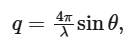
(2.1)
where λ is the wavelength of the incident X-ray beam, and 2θ is the scattering angle. Scattering patterns were plotted in Origin2021. Area under the curve (AUC) was calculated over an equivalent q interval for each set of profiles using Origin2021 and is the region enclosed by the scattering profile and the line drawn between qmin and qmax on the curve.
2.3.2. Monoolein cubosomes
MO cubosomes (500 μl) were mixed with S. aureus cell-free supernatant (500 μl) via pipette, dispensed into 1.5 mm glass capillaries for SAXS (Capillary Tube Supplies Ltd, Cornwall, UK) and flame-sealed. SAXS patterns were obtained on a point-collimated Bruker NanoSTAR II instrument with TXS rotating Anode (Cu Kα X-ray source; 8.04 keV) equipped with a Vantec-2000 2D detector (Bruker AXS, WI, USA) (beam ⌀ = 1 mm (full width half maximum); Φ = approx. 45 million cps; available q-range 0.01 Å−1 to 0.35 Å−1) and temperature-controlled stage (±0.1°C). SAXS capillaries were incubated at 37°C throughout measurement. Detector images were radially integrated using the Bruker SAXS software package. Liquid crystal phases were identified using Bragg peak q-spacing as described by Hyde (see supplementary material, §S3.1, for further details) [34–36].
2.4. Real-time quantitative polymerase chain reation
2.4.1. Materials
RNA-free water and RNAse Zap were purchased from Thermo Fisher. The RNA Miniprep Kit and QuantiNova RT Kit were purchased from ZymoBIOMICS and QIAGEN, respectively. SYBR Green mastermix was purchased from Invitrogen.
2.4.2. Cell culturing and RNA extraction
Staphylococcus aureus culture in MH broth was prepared as previously outlined, streaked onto agar and incubated overnight (37°C). Single colonies were selected to inoculate sterile MH media in triplicate. Each replicate culture was incubated (37°C, 4 hours), separated into two equal volumes, diluted 1 : 1 (v : v) with fresh MH media or MO cubosomes (50 mg ml−1 in PBS), and incubated for a further 2 hours. Cells were pelleted by centrifugation (10 000 × g, 5 min, 4°C), the supernatant discarded, and cells were stored at −80°C overnight. For all subsequent procedures, samples were kept on ice unless otherwise stated, and RNaseZap was used to decontaminate surfaces. The cells were redispersed in lysis buffer using vortex mixing (VELP, Italy) and lysed using a bead mill homogenizer (4.0 m s−1, 30 s; Bead Ruptor Elite, OMNI International). RNA was extracted and purified using an RNA Miniprep Kit.
2.4.3. Real-time quantitative polymerase chain reation
Sample RNA concentrations were determined by absorbance at 260 nm (Nanodrop one, Thermo Fisher) and diluted to 1 μg ml−1 with RNase-free water. Reverse transcription (RT) was performed using a QuantiNova RT kit and thermocycler (T100 Thermal Cycler, Bio-Rad) to afford complementary DNA (cDNA).
Quantitative polymerase chain reation (qPCR) was conducted using SYBR Green mastermix (Mic qPCR Cycler, Biomolecular Systems) and analysed using micPCR 2.8.1. Cubosome particle size, oligonucleotide primer sequences and qPCR results are listed in supplementary material, tables S1–S3.
3. Results and discussion
3.1. Microbial digestion of phospholipid liposomes
Staphylococcus aureus and P. aeruginosa (PA01) were studied as clinically relevant opportunistic pathogens that commonly colonize the human body and are implicated in a wide range of infections. Both S. aureus and P. aeruginosa have been targeted by lipid-based antimicrobial formulations in previous studies, highlighting both the clinical need for more effective antimicrobial agents against these pathogens and the growing interest in lipidic delivery systems for their treatment [27,37–39]. They both express a variety of substrate-specific and context-dependent lipases, often relying on environmental cues for activation [40,41]. Pseudomonas aeruginosa is particularly geared towards phospholipid digestion for energy harvest, expresses phosphatidylethanolamine (PE)- and phosphatidylcholine (PC)-specific phospholipases PlcB and PlcH, and demonstrates chemotaxis towards phospholipids [42–44]. Staphylococcus aureus preferentially incorporates exogenous fatty acids into its cell membrane and primarily uses phosphatidylglycerol, lysyl-phosphatidylglycerol and cardiolipin as its major membrane phospholipids and is geared more towards the digestion and use of these lipids rather than PE and PC [45–47]. Moreover, the strain of S. aureus studied in this work expresses lip1 and lip2, which encode glycerol-ester-specific lipases (supplementary material, figure S1; further discussed in supplementary material, §S4). These species-specific subtleties in lipase substrate specificity are demonstrated in the digestion of DOPE/DOPC liposomes with cell-free S. aureus and P. aeruginosa culture supernatants (figure 2), which consists of all expressed extracellular proteins and vesicles (⌀ < 450 nm).
Figure 2.
Phospholipid digestion with all enzymes expressed by live cultures. (a–d) Stacked SAXS profiles for DOPE/DOPC liposomes stabilized with (a,b) F127 or (c,d) DSPE-PEG-2000 over the course of digestion with cell-free culture supernatants from (a,c) S. aureus and (b,d) P. aeruginosa PA01. Scattering profiles remain unchanged, except for F127-stabilized liposomes with cell-free culture supernatant from P. aeruginosa PA01. (b) The loss of scattering intensity over time signifies fewer lipid nanoparticles in the beam path to interact with. (e) AUC of background-subtracted SAXS patterns (a–d) expressed as a percentage of the first time point. AUC was calculated over an equivalent q interval for each set of profiles and is the region enclosed by the scattering profile and the line drawn between qmin and qmax. Only phospholipid liposomes stabilized with F127 were digested by P. aeruginosa (b) as indicated by the decrease in AUC over time.
Consistent with the preferential activity of S. aureus lipases, both liposomal formulations exposed to S. aureus culture supernatant remained unchanged over 14 hours (figure 2a,c). In the presence of P. aeruginosa culture supernatant, however, liposomes stabilized with Pluronic F127 exhibited a gradual reduction in the scattering intensity of the broad lamellar peaks, indicative of progressive digestion (figure 2b). Liposomes stabilized with DSPE-PEG-2000 remained unchanged over time (figure 2d).
The digestion of PE and PC assemblies by phospholipases has been well-characterized in the literature (supplementary material, figure S2) [22]. During digestion, PE assemblies retain diacylglycerol (DAG) digestion products and consequently adopt an H2 structure. Conversely, PC assemblies do not retain DAG digestion products, which consequently oil out. Homogenizing via probe sonication temporarily adopts the H2 structure. This demonstrates that both PE and PC assemblies adopt an H2 structure when phospholipase C digestion products are retained in the assembly. PE assemblies retain DAG via H-bonding of NH3+ moieties. PC assemblies do not retain DAG as they lack H-bonding partners. Based on this literature, it is unclear whether the 1 : 1 molar ratio of PE : PC studied here retains DAG during digestion, however, can be identified by the formation of H2 Bragg peaks.
The Bragg peaks that would indicate the formation of non-lamellar phases are not present in the scattering patterns reported in figure 2. This indicates that a 1 : 1 molar ratio of DOPE : DOPC provides insufficient H-bonding to retain DAG in the assembly. Further, the loss of scattering intensity over time signifies a reduction in the concentration of liposomes over time, consistent with the loss and phase separation of DAG from the liposomes.
The differences in stabilizing effect provided by F127 and DSPE-PEG-2000 are attributed to surface chemistry at the particle–fluid interface. Lipases must adsorb onto hydrophobic surfaces at the oil–water interface to engage the molecular substrate [48]. The steric stabilizer layer lies at the interface between the surface of the nanoparticle and the external aqueous environment, acting as the mediator between these two domains. Polymer chain length, conformation and hydrophobicity/hydrophilicity all influence whether a molecule has continued access to the particle surface [18]. Bilayer-anchored stabilizers (e.g. DSPE-PEG-2000) maintain a consistent steric barrier, whereas surface-adhering stabilizers (e.g. F127) are labile—their detachment enables enzymes access to the LNP surface (figure 2e) [19,48].
3.2. DSPE-PEG-2000 accelerates enzymatic phase transitions in monoolein cubosomes
To determine whether bilayer-anchored stabilizers confer greater protection against bacterial lipases for other classes of LNPs, monoolein cubosomes were prepared with Pluronic F127 alone or in combination with DSPE-PEG-2000.
3.2.1. Formulation development and phase behaviour
A series of PEGylated cubosome formulations with varying DSPE-PEG-2000 content (1–9 wt%) were screened for colloidal stability (supplementary material, figure S3, tables S4 and S5). The formulation containing 5 wt% F127 + 5 wt% DSPE-PEG-2000 demonstrated optimal stability over 1 week in terms of hydrodynamic diameter (150–300 nm) and polydispersity index (<0.2) and was therefore selected for enzymatic digestion studies. Notably, the addition of DSPE-PEG-2000 consistently led to a shift towards lower q values and broader Bragg peaks compared with F127-only formulations (supplementary material, figure S4 and table S6). This increase in lattice parameter corresponds to lattice swelling (approx. 10 Å increase), while the peak broadening reflects increased lattice heterogeneity and disorder. These observations suggest that a fraction of DSPE-PEG-2000 is incorporated into the particle's internal nanostructure, with internalized PEG chains occupying the water channels and encouraging swelling. Bragg peak broadening suggests this incorporation is probably non-uniform throughout the internal matrix. Such heterogeneity may arise from physical confinement, resulting in localized regions of higher PEG chain density and consequently greater water-channel swelling towards the particle's surface as well as regions of lower PEG chain density within the water channels closer to the particle's centre, where PEG confinement would be energetically unfavourable.
Phase behaviour (supplementary material, figure S5 and table S7) for the MO/OA/F127/DSPE-PEG-2000 system confirmed identical liquid crystalline phase behaviour to the MO/OA/F127 system previously reported [49], with reorganization of the nanostructure towards increasingly curved bilayer structures observed as OA content increased (Im3m→H2→Fd3m). Notably, the concentration of OA required to achieve transformation to H2 and Fd3m phases was not affected by the addition of DSPE-PEG-2000.
The disruptive effect of DSPE-PEG-2000 on non-lamellar structures became even more apparent when attempting to prepare cubosomes using DSPE-PEG-2000 as the sole stabilizer. At concentrations up to 9 wt%, rapid aggregation occurred upon dispersion despite probe sonication. At 29 wt% (5 mol%) DSPE-PEG-2000, dispersion was achieved but resulted in large, polydisperse vesicles with complete loss of cubic symmetry (supplementary material, figure S6). Indeed, there are no reports of monoolein cubosomes stabilized solely with DSPE-PEG-2000, and only sparse literature describing dual F127/DSPE-PEG-2000 stabilization [50–53].
The physical basis for this incompatibility lies in the geometric frustration imposed by lipid-PEG anchoring. In cubic phases, the lipid bilayer curves in three dimensions to form interconnected water channels. Molecules must occupy the internal water channels or extend into the external aqueous medium. With cubosomes having significantly greater internal surface area than liposomes, a significantly greater proportion of PEG chains are internalized rather than externalized. Internalized PEG chains create osmotic/steric pressure, which opposes the natural curvature of the lipid bilayer and results in lattice swelling. Similar observations have been reported for phytantriol/DSPE-PEG/F127 cubosomes, noting that higher lipopolymer concentration and longer PEG chains both increase the lattice swelling effect [54]. At 5 mol% DSPE-PEG-2000, corresponding to 20 lipids per PEG chain (equivalent to PEGylated liposomes), this internal osmotic/steric pressure is too great for the cubic structure to form. In theory, a much higher lipid : PEG chain ratio would be necessary for cubosomes to achieve the same level of surface coverage that is achieved with liposomes. Therefore, it may not be possible to adequately stabilize non-lamellar structures using bilayer-anchored stabilizers owing to the internal sequestration of polymer chains resulting from their significant internal bilayer surface area.
A long-standing goal in drug delivery is to create PEGylated liposomes that remain stable during circulation but undergo structure-mediated drug release upon encountering disease-specific stimuli (e.g. bacterial lipases, low pH or specific biomolecules). Our analysis demonstrates that bilayer-anchored stabilizers fundamentally oppose the lamellar→non-lamellar phase transitions required for such responsiveness owing to the osmotic/steric pressure created by internalized polymer chains. PEG chains shorter than 2000 Da offer less protection against protein attachment [18]. Moreover, the tendency for PEG chains to become internalized rather than externalized in non-lamellar assemblies leads to poor surface coverage. Together, this suggests that bilayer-anchored stabilizers may not be suited for designing stimuli-responsive liposomes that undergo structural reorganization to non-lamellar assemblies.
3.2.2. Enzymatic digestion: counterintuitive acceleration of phase transitions
Our previous work has shown that S. aureus causes monoolein cubosomes to undergo structural reorganization towards increasingly curved bilayer structures consistent with substrate-specific lipase activity (Im3m→H2→Fd3m) [15]. To probe whether different steric stabilizers influenced S. aureus lipase activity, cubosomes and PEGylated cubosomes were subjected to digestion. We hypothesized that DSPE-PEG-2000 would sterically hinder lipase access and slow enzymatic digestion; however, PEGylated cubosomes underwent phase transitions earlier and faster than F127-only cubosomes (figure 3).
Figure 3.
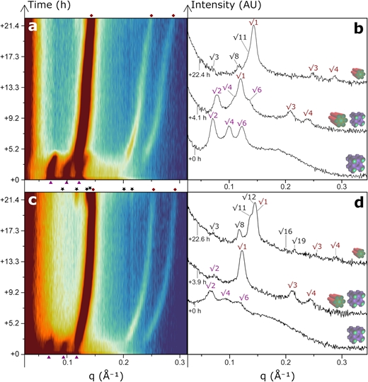
Digestion of (a,b) monoolein cubosomes and (c,d) PEGylated cubosomes with all enzymes expressed by S. aureus. Symbols indicate Bragg peaks; colour denotes liquid crystal structure. (a) MO/F127 cubosomes transition from Im3m (▲ √2, √4, √6) to H2 (◆ √1, √3, √4) space group at t = 3.1–4.6 hours. The H2 lattice gradually shrinks over the course of digestion (approx. 10 Å). (b) Individual frames at key timepoints reveal the beginning of transition to Fd3m (★ √3, √8, √11) with most identifying peaks eclipsed by overlapping H2 peaks. (c,d) With 5 wt% DSPE-PEG-2000, Im3m Bragg peaks are broader and observed at lower q, consistent with inhomogeneous swelling of the internal water channel network. The Im3m→H2 transition begins earlier at t = 2.4–3.4 hours, and H2→Fd3m from t = 20.6 hours (★ √3, √8, √11…).
For F127-stabilized cubosomes, the Im3m→H2 transition occurred between t = 3.1–4.6 hours, with the emergence of Fd3m Bragg peaks predominantly obscured by overlapping H2 peaks (figure 3a,b). For PEGylated cubosomes, the Im3m→H2 transition occurred between t = 2.4–3.4 hours, and clear Fd3m peaks appeared by t = 20.6 hours (figure 3c,d). Given that DSPE-PEG-2000 prevented digestion for phospholipid liposomes, the accelerated digestion observed with PEGylated cubosomes seems paradoxical.
Considering the geometry of the system, the presence of DSPE-PEG-2000 is expected to promote more positively curved bilayer structures [55]. Internalized PEG chains increase the osmotic pressure within the internal water channels, leading to lattice swelling at 5 wt% DSPE-PEG-2000 (supplementary material, table S6), and the transition to lamellar structure at 29 wt% (supplementary material, figure S6) [56]. These effects induced by DSPE-PEG-2000 are expected to present an even greater degree of frustration for the H2 lattice (supplementary material, table S8) given that the diameter of the water channel in the initial Im3m cubosome (53 Å) decreased by greater than 40% when the phase transition occurred during digestion (30 Å) (assuming a constant average lipid chain length of 15 Å, calculations in supplementary material, §S3.2) [35,36]. Hence, the OA produced during digestion had a much greater influence on liquid crystal phase behaviour by partitioning to hydrophobic ‘void’ regions and reducing the energy cost associated with H2 phase formation than the opposing osmotic pressure associated with internalized PEG chains.
Moreover, the internalization of stabilizer PEG chains leaves the primary stabilizing role to be performed by F127. Cubosomes containing DSPE-PEG-2000 (⌀ ∼ 184 nm) are only slightly larger than those stabilized with F127 alone (⌀ ∼ 156 nm) (supplementary material, table S6); if particle size is influencing digestion kinetics, it is slightly enhancing the digestion of F127 cubosomes. Phase-behaviour studies of the MO/OA/F127/DSPE-PEG-2000 system here and MO/OA/F127 systems reported in literature [49] show that the Im3m→H2→Fd3m transitions occur at equivalent OA concentrations, indicating that the amount of OA required to elicit phase transformation is equivalent for cubosomes and PEGylated cubosomes.
This acceleration in kinetics is therefore rationalized as a consequence of swelling the cubosome lattice, thereby increasing the area per lipid molecule and consequently improving the accessibility of digestive enzymes to lipid headgroups [48,57–59].
4. Conclusions
In this study, phospholipid liposomes and monoolein cubosomes were enzymatically digested by live bacteria to compare the stabilizing effect provided by bilayer-anchored and surface-attached steric stabilizers. The surface-attached stabilizer, Pluronic F127, failed to adequately protect both LNPs from enzymatic digestion. The bilayer-anchored stabilizer, DSPE-PEG-2000, was found to inhibit the digestion of liposomes yet accelerate the digestion of cubosomes. The accelerating effect was attributed to lattice swelling, increasing the area per lipid molecule and promoting access of the enzyme to constituent lipids. Together, these results demonstrate that particle nanostructure and stabilizer mode of attachment play key roles in determining whether steric stabilizers provide an inhibitory or accelerating effect for the digestion of LNPs.
Most importantly, steric stabilizing agents do not offer unconditional protection for LNPs against enzymatic attack. The steric stabilizer mediates the interactions between the particle and the surrounding environment. Understanding this behaviour in non-lamellar assemblies can help facilitate the development of stimuli-responsive LNPs with greater selectivity and control over the biological stimuli it responds to under diverse physiological conditions.
View further article information including Ethics, Data accessibility, Declaration of AI use, Authors' contributions, Conflict of interest declaration, Funding, Theme, Acknowledgements, and References here: www.bit.ly/RSFS162
#Feature #ChemistryArticle #AustralianAuthoredContent